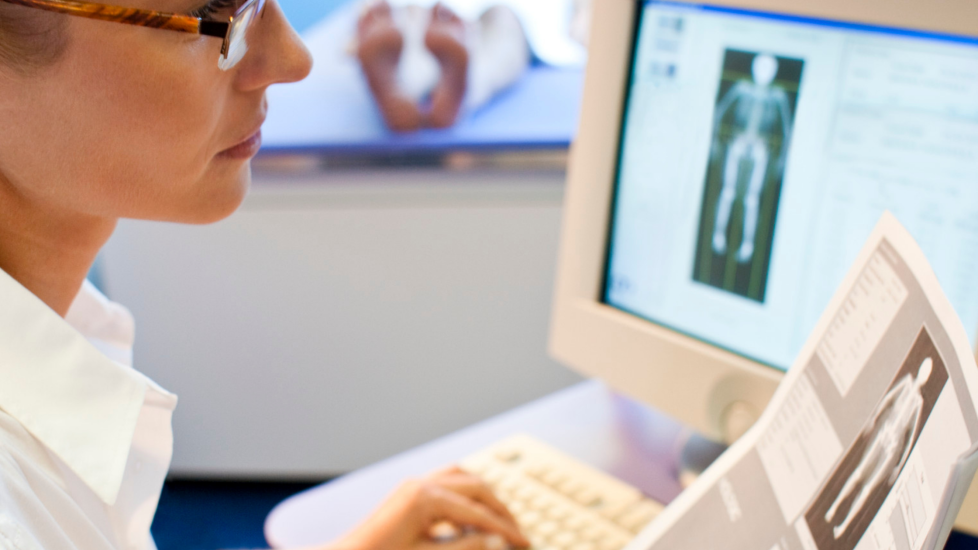
Bone Density Scans or Dual Energy X-Ray Absorptiometry (DEXA) scans, use a low dose of X-ray to establish the density (or strength) of bones.
Bone density scans are used to diagnose or assess the risk of osteoporosis, a condition that makes bones weak and likely to break (fracture). Osteoporosis is a result of the body not making enough bone, losing too much bone or a combination of both. Bone density does reduce as part of the normal course of ageing and commonly affects post menopausal women, due to reduced oestrogen levels. However, Osteoporosis can also be attributed to:
(NHS.org.U.K.)
Bone metastasis can also increase risk of serious bone events including fracture and DEXA scans are sometimes used to establish fracture risk for patients with bone metastasis.
DEXA scans are more reliable than standard x-rays in identifying bones with reduced density and identifying fracture risk and use a lower dose of X-ray. The scan works by passing x-rays through bones and measures how much radiation from the x-ray is passed through and how much is absorbed.
DEXA scans are quick and painless, and the Radiographer will often stay in the room with you.
“It has been a number of years since I had a bone density scan but I do remember it being a very straightforward experience. I changed into a hospital gown as there should be no metal present for the scan. I was asked to lie flat on the bed under the scanner and the scanner was moved into place above me. The radiographer remained in the room with me and it took less than 5 minutes to complete the scan. Results were then sent back to my medical team.”
Contributions from Sarah & Claire, members of the Make 2nds Count community.