9th February 2021 Scans

An ultrasound scan is a diagnostic procedure that creates an image from a body part inside the body from high-frequency sound waves. Ultrasound scans don’t have any known risks like other imaging examinations as there is no exposure to radiation.
In oncology an ultrasound scan can be used to aid diagnosis of a condition and guide surgeons during certain procedures. Some types of ultrasound scan require some preparation before in order to optimise the quality of the scan. This may be to have a full bladder, or to not eat for a period before the scan.
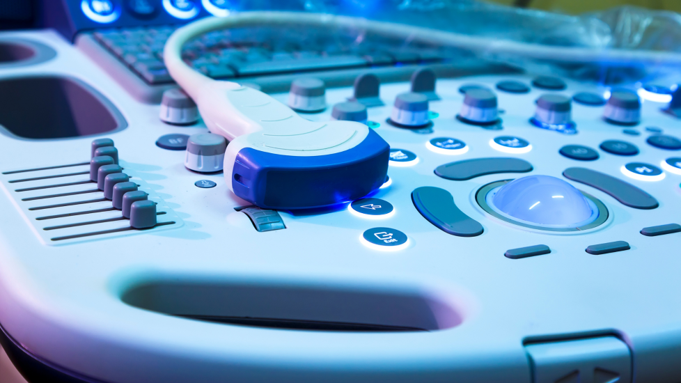
During the procedure, a sonographer or radiologist uses a small device called an ultrasound probe over the area of the body that is to be imaged. The probe gives off high-frequency sound waves (which cannot be heard) over that area and the waves are echoed back off the surrounding body tissues to the probe which then creates an image in the monitor. An ultrasound scan can be external, internal or as an endoscope to reach further into the body.
An external ultrasound scan is non invasive. Gel is placed on the probe, which is then placed on the skin and moved over the area. This is generally very comfortable, the gel can be a bit cold and there may be some discomfort if pressure needs to be applied to enhance the quality of the image. This is commonly used over a breast lump and liver in breast cancer patients.
An internal ultrasound is most commonly used to look at the ovaries or womb in women and prostate in men.
An endoscopic ultrasound is often used to assess the stomach and oesophagus. The probe is attached to a long flexible tube and passed down the throat. This can be uncomfortable, but a sedative is normally given, through a needle in the hand.
The sonographer or radiologist may tell you the results of the scan immediately, but it is common for the images to be analysed and a report sent to the doctor who requested the scan to then feedback the results at another appointment.
My experience of an Ultrasound
I have had 3 ultrasound scans in the past year.
They are non invasive and generally you don’t need to do anything in preparation ( if you do ,you will be advised beforehand). I do tend to wear clothes that are easy to get on and off to save time! Sometimes you are asked to wear a gown but have noticed since Covid reared its ugly head this hasn’t happened.
The sonographer and assistant are in the room and the lights dimmed . The jelly is then applied to the area (sometimes if you are lucky they will warm it up first!) . The probe is then moved over the area they are looking at. It is not painful but may be uncomfortable when they need to apply pressure. I have also had guided biopsies when they use the ultrasound to make sure they are taking the biopsy from the correct place. These may or may not involve local anaesthetic to the area . I have had both , but even without anaesthetic it wasn’t too painful.
After the scan you are given tissue to clean yourself. I was told the results straight away (apart from the guided biopsy to my sternum as that needed to be tested) .
Out of all the scans I have had I find the ultrasound the easiest to deal with .
Contributions by Carol & Sarah,
Make 2nds Count Community Members